


Osteochondrosis of the cervical spine (CS) is one of the most common pathologies of the musculoskeletal system. Every year doctors diagnose this disease more and more often, and its course becomes more serious. According to statistics, in women degenerative-dystrophic changes in the upper part of the spine occur more often, especially in postmenopausal patients. The main symptoms of cervical osteochondrosis in women are pain, limited mobility and cerebrovascular insufficiency, and this is dangerous not only for health, but also for life. To protect yourself from the dangerous consequences of the pathology, it is necessary to start treatment in the initial stages. It is important to conduct complex therapy and lifestyle changes to stop the destruction of spinal segments and prevent serious complications.
Development of the disease
The cervical spine is most vulnerable to various injuries and degenerative changes. This is due to the fact that this segment is the most mobile and the muscles here are weak. Small cervical vertebrae bear heavy loads every day, which leads to the gradual destruction of the intervertebral discs. The vertebrae put pressure on each other, which causes the cartilaginous pads between them to leak a lot of fluid and begin to degrade and deform.
Also, osteochondrosis of the cervical spine develops due to insufficient nutrition of the cartilaginous tissue. And the spinal canal in this area is narrow, so it is often compressed, causing neurological symptoms.
Pathology in women in the initial stages is manifested by heaviness in the back of the head, tingling in the hands, etc. Patients often confuse the first signs of the disease with fatigue.
In the neck area there are numerous blood vessels and nerve roots; neurological disorders may also occur when they are compressed. It is especially dangerous if a deformed disc or vertebra compresses the vertebral artery, which supplies important parts of the brain. When compressed, coordination of movements is impaired, a woman may lose balance, vision and hearing deteriorate, and the risk of stroke increases.
Reference.According to statistics, most often cervical osteochondrosis is found in patients aged 25 to 40 years. This is due to a massive decrease in physical activity and sedentary work. Women are diagnosed with the disease more often than men, as they have more fragile vertebrae and thin bone tissue.
Doctors distinguish 4 stages of osteochondrosis of the spine:
- Phase 1– the intervertebral disc loses part of its moisture, its height decreases and cracks may appear on the fibrous ring (outer shell). This is the stage of cervical chondrosis, which is difficult to identify because it presents unexpressed symptoms. The neck gets tired quickly, there is discomfort, heaviness in the damaged area, sometimes there is a slight pain that quickly passes.
- Phase 2– cracks on the surface of the disc increase, the nucleus pulposus (the gelatinous contents of the disc) shifts and may protrude through the damaged areas. This is what protrusions of the cartilaginous covering appear that can compress the spinal cord and its roots. Severe pain, weakness, limited mobility appear periodically, and numbness of the face, neck, shoulders and arms may occur.
- Phase 3– the protrusion breaks through the outer shell of the disc, thus forming a hernia. The pain becomes more pronounced and neurological disorders are present.
- Phase 4– the disc is almost completely destroyed, the vertebrae rub against each other and bony growths (osteophytes) appear on the edges which have the purpose of stabilizing the damaged segment. Nerve endings, spinal cord and blood vessels are damaged. Adjacent joints begin to be damaged. The clinical signs are pronounced.
It is easier to stop degenerative-dystrophic changes in the first two stages of osteochondrosis of the spine. In phase 3, comprehensive treatment will help stop further destruction of the spinal segment. In the last stage, surgery cannot be avoided.
Causes
Osteochondrosis of the spine is a complex and long process, which most often has several causes. In most cases, the pathology occurs due to a sedentary lifestyle, poor nutrition and metabolic disorders. The disease often occurs due to injuries or due to the natural aging of the body and the weakening of its defenses.
Doctors identify the main causes of osteochondrosis of the spine in women:
- Violation of metabolic processes.
- Passive lifestyle.
- Genetic predisposition.
- Chronic muscle tension around the cervical segment.
- Postural distortion.
- Deficiency of fluids and nutrients in the body.
- Prolonged stay in an uncomfortable position (neck stretched forward and back curved).
- Overweight.
- Frequent use of high-heeled shoes.
- BUY injuries.
- Lifting heavy objects.
- Autoimmune diseases.
- Frequent stress, chronic fatigue.
- Hypothermia.
- Infectious diseases.
- Neck too long or short, etc.
All these factors cause malnutrition of the intervertebral discs and lead to their degeneration.
Female cervical osteochondrosis can be caused by pathologies of the vertebral artery associated with genetic predisposition, intrauterine disorders and injuries during childbirth. The disease can occur due to rheumatism, endocrine disorders, excessive load on the cervical segment during pregnancy and local overload.
Important.The main cause of cervical osteochondrosis in women is menopause, as well as the changes associated with this period. At this stage, the concentration of progesterone in the body, which is very important for bone tissue, decreases. The likelihood of degenerative changes is associated with age-related weakening of the neck muscles and weakening of spinal support in this area.
Symptoms
Osteochondrosis is characterized by a wave-like course, when the acute period is replaced by remission. Exacerbation can be caused by infection, injury, hypothermia and prolonged tension on the neck.

The first signs of cervical osteochondrosis in women are headache, discomfort and heaviness in the neck. It is important to distinguish pain due to chondrosis from migraine or autonomic dysfunction over time.
Clinical manifestations of osteochondrosis of the spine in women are caused by neurological syndromes:
- Cervical dysscalgia occurs when nerve endings are irritated by fragments of damaged cartilage lining. Then a specific creaking in the neck appears, a pain that becomes more pronounced when moving the head and after sleep.
- Scalenus syndrome is a consequence of damage to the vessels and nerves of the brachial plexus and subclavian artery. This complex of symptoms is accompanied by pain from the inner surface of the shoulder to the hand on the injured side. The limb becomes pale, cold, swollen, and numbness occurs. Neck pain extends to the back of the head when the patient turns his head.
- Humeral periarthrosis syndrome: Dystrophic changes affect the tendon fibers surrounding the shoulder. Painful sensations from the neck radiate to the shoulder and shoulder girdle. There is a forced position of the neck: it is tilted to the affected side, and the shoulder is slightly lowered.
- Vertebral artery syndrome: A blood vessel is compressed by fragments of a damaged disc or by osteophytes (depending on the stage of the disease). The patient feels dizzy and has headache, nausea and sometimes vomiting. The pain is localized in the back of the head, crown and temples.
- Cardiac: The nerve bundles of the spinal cord are damaged. Heart pain and arrhythmia occur. If C3 is damaged, pain appears in the middle of the neck, the tongue swells, and the patient cannot chew food normally. If C4 is injured, discomfort appears in the area of the shoulder girdle, collarbone and heart. When C5 is affected, the painful reaction from the neck spreads to the shoulder girdle, the inner surface of the shoulder. C6 irritation causes pain from the neck and shoulder blade to the shoulder girdle and spreads up the entire arm to the thumb. If C7 is damaged, the pain syndrome spreads to the back of the shoulder girdle, affecting the entire hand, including the index and middle fingers. When C8 is compressed, the pain spreads from the affected area to the elbow and little finger.
In addition, a woman's emotional sphere may be disturbed, weakness may occur, she becomes anxious and touchy. Insomnia often occurs, memory and attention are weakened due to regular headaches.
Symptoms of a cerebrovascular accident occur when a woman suddenly throws her head back, tilts it, or does work that puts stress on her arms and cervical spine, such as when she digs, paints a ceiling, or carries heavy objects.
Poor cerebral circulation manifests itself with dizziness, unsteady gait, spots before the eyes, tinnitus, weakness and nausea. In some patients, the voice becomes hoarse, sometimes disappears and a sore throat appears.
Osteochondrosis during menopause is accompanied by migraines, increased body sweating in the area between the neck and shoulder girdle. When the vertebral artery is compressed, the functioning of the cardiovascular system is disrupted.
If the disease lasts a long time, circulatory failure occurs in important centers that perform neuroendocrine functions. Due to the increased permeability of the vascular walls, atherosclerosis of the cerebral and cardiac arteries develops.
Establish the diagnosis
If you notice symptoms of osteochondrosis, go to a therapist. After a visual examination, the specialist will refer you to an orthopedist, vertebrologist or neurologist.
The following methods are used to diagnose cervical osteochondrosis:
- X-ray allows us to find out that the patient's vertebrae are displaced, there are osteophytes on their edges, the distance between the vertebrae has decreased, etc. For this reason, the study is carried out on different levels. To detail the characteristic changes, the doctor takes targeted photographs.
- CT scan of the cervical spine provides detailed information on pathological changes in the vertebrae. This method allows you to obtain three-dimensional images for more detailed study; it is used in severe diagnostic cases.
- MRI is used to accurately evaluate the condition of the soft tissues (nerves, blood vessels, ligaments, muscles) in the affected area.
- Electromyography allows you to verify the conductivity of the nerve fiber.
Doctors may also prescribe an ultrasound (Doppler ultrasound of the main arteries of the brain) to determine the state of blood flow in this area.
Conservative treatment
In the initial stages, treatment of osteochondrosis of the spine in women can be carried out at home. However, a doctor must come up with a treatment regimen. It is important to understand that this is a long process and a full recovery is unlikely to be possible (especially for older women).
Complex treatment includes:
- Taking medications.
- Use of orthopedic devices.
- Physiotherapy.
- Physiotherapy procedures.
- Massage, manual influence.
- Alternative treatments.
Conservative methods will help relieve pain, inflammation, normalize muscle tone, improve metabolic processes, nutrition of damaged segments of the spine, etc. With timely therapy, it is possible to stop pathological changes.

Treatment of cervical osteochondrosis in women is carried out with the use of drugs that will help improve the metabolism of the cartilaginous pads between the vertebrae, relieve inflammation and pain. For this purpose, the following drugs are used:
- NSAIDs. They will help relieve inflammation and pain of mild to moderate severity.
- Analgesics. Relieves pain.
- Drugs to improve cerebral circulation.
- Muscle relaxants help relieve muscle spasms.
- Chondroprotectors. They help stop disc destruction, improve metabolic processes and speed up recovery.
- Magnesium-based medicines.
- Nootropics. They stimulate brain functioning by normalizing blood circulation and have a mild sedative effect.
Reference.In case of severe pain that is not relieved by oral medications, therapeutic blockades are used, for example with an anesthetic solution or NSAIDs.
The treatment can be supplemented with anti-inflammatories and painkillers in the form of gels, creams and ointments. They will be effective in the remission phase or in combination with oral drugs.
The decision on the choice of drug combinations is made by the doctor. The specialist will draw up a treatment regimen and also determine its dosage. It is important to follow his recommendations, since many of the drugs described above can lead to dangerous complications.
During the acute phase of osteochondrosis of the spine, a woman should refuse strenuous physical activity. To relieve the cervical segment, it is necessary to wear a special corset (Schants collar), which will fix the vertebrae in the correct position. This device is recommended for use during prolonged sedentary work or heavy physical work.
Physiotherapeutic procedures will help relieve pain and improve blood circulation in the damaged area:
- Diadynamic therapy.
- Magnetotherapy.
- Electrophoresis with an anesthetic, glucocorticosteroid, proteolytic agent.
- Electroanalgesia.
- Ultraviolet irradiation, etc.
The therapeutic effect appears approximately after the third session, then headaches, hearing and vision disorders, dizziness weaken or disappear, sleep normalizes and the general condition improves.
Using underwater traction of the cervical segment, it is possible to expand the distance between the vertebrae, free a nerve or blood vessel from compression, and restore the normal position of the vertebrae.
Massage will normalize muscle tone and reduce the flow of lymphatic fluid, which causes swelling. After several sessions, blood circulation in the damaged area improves.
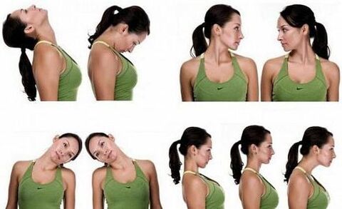
Therapeutic gymnastics is one of the most effective methods of treating osteochondrosis of the spine. Physical therapy allows you to strengthen weak neck muscles, which will then take on some of the load from the spine and help stop or slow down degenerative changes. During exercise, blood circulation improves, metabolic processes and disk nutrition are accelerated, which has a positive effect on their condition.
Women should exercise every day. They consist of simple but effective exercises. The complex consists of turns, tilts of the head in different directions, as well as neck movements, during which the arms are used. These items can be performed at home, but only after permission from a doctor. Physical therapy is performed only in the remission phase.
Complex treatment can be supplemented with reflexology (acupuncture), hirudotherapy (treatment with leeches), swimming, etc.
Surgery
The operation is prescribed in the last stages of osteochondrosis of the spinal cord, which are accompanied by severe destruction of osteochondral structures. Furthermore, surgery cannot be avoided if conservative methods are ineffective or if the spinal canal has narrowed significantly.
In the cases indicated above, an anterior cervical discectomy is performed. During the procedure, the doctor immobilizes the damaged segment of the spine and removes the hernia that was compressing the spinal nerve. Then the vertebrae between which the disc was removed are fused. If necessary, the space between the vertebrae is filled with a synthetic insert (cage).
After 3-5 days the patient is discharged home. The rehabilitation period is approximately 12 weeks. To speed up recovery, you need to take medications, wear a corset, lead a healthy lifestyle, undergo physiotherapeutic procedures and possibly perform physical therapy.
Lifestyle recommendations
To quickly eliminate the unpleasant symptoms of osteochondrosis and stop degenerative-dystrophic changes in the cervical segment, you need to adjust your lifestyle. To do this, the patient must follow these recommendations:
- Take walks every day, avoid running, jumping and other explosive activities.
- Do not carry heavy objects.
- You can not sit for a long time, in extreme cases, wear a corset and periodically take a horizontal position.
- Perform special physical exercises for the back muscles at home.
- Sleep on an orthopedic mattress and a special pillow.
- Follow a diet, replenish your diet with foods rich in magnesium, calcium (nuts, dairy products, seafood, legumes), as well as plant fiber, chondroitin (jellied meat, gelatin). Avoid fatty, fried, too salty foods and alcohol. Your doctor will advise you in more detail about nutritional rules. But in any case it must be correct.
Hypothermia should not be allowed; warming will be useful in the absence of an inflammatory process.
Complications
In the absence of timely treatment for cervical osteochondrosis, a woman may experience the following consequences of the pathology:
- The probability of a protrusion, which after some time turns into a hernia. The swelling compresses the spinal cord and its nerves, causing neurological disorders.
- Osteophytes appear when the disc is severely damaged and irritate the spinal nerves and blood vessels.
- In advanced cases, severe weakening of the neck muscles or incomplete paralysis is possible, so the head involuntarily hangs to the side or forward.
- Compression of the vertebral arteries, reduced circulation in the affected area. This condition can cause neuralgia (pain along the nerve), hearing and vision problems.
- Paralysis (incomplete or complete) of the hands.
- Brain stroke, etc.
If a woman copes with the problem in the early stages of osteochondrosis of the spinal cord, she will be able to prevent the conditions described above.
Preventive measures
Ideally, prevention of osteochondrosis of the spine should be carried out during the period of intrauterine development. The expectant mother must exclude factors that negatively affect the development of the fetus: infections, oxygen starvation, intoxication. If there has been an injury at birth, the newborn must undergo treatment.
To reduce the likelihood of developing osteochondrosis of the spine, a woman should follow these recommendations:
- Load the spine evenly, for example carry a load with both hands or alternately with the right and then the left.
- Don't lift too much weight alone.
- Try to avoid neck injuries and hypothermia.
- While working in the vegetable gardens, take a break every 1. 5 hours and lie down to rest for 20 minutes.
- Choose shoes with elastic soles that cushion impacts when running or jumping.
- When sitting for long periods of time, use a high-backed chair and a headrest or wear a corset.
It is also important to eat well, control your weight, avoid stress, take vitamin supplements for medical reasons and promptly treat pathologies that can cause osteochondrosis. During the remission phase, it is recommended to visit sanatoriums to undergo a course of treatment.
The most important
As you can see, osteochondrosis of the cervical spine occurs more often in women than in men, since the former have more fragile vertebrae and thin bone tissue. Patients during the postmenopausal period are particularly susceptible to the pathology. The disease manifests itself with pain, neurological disorders and dangerous symptoms of cerebrovascular accident. It is recommended to start treatment in the initial stages to avoid dangerous complications of osteochondrosis. To do this, a woman must take medications, adjust her lifestyle, attend physiotherapeutic procedures, massage, do physical therapy, etc. Surgical treatment is indicated only in advanced cases. To prevent pathology, it is necessary to maintain moderate physical activity, timely treat injuries and diseases that can provoke osteochondrosis, etc.